Best 3D-4D Sonography Centre in Ahmedabad
3D/4D sonography offers detailed images of the fetus during pregnancy, providing a unique and emotional experience for expectant parents.
3D/4D Sonography at Prabhu Women’s Hospital
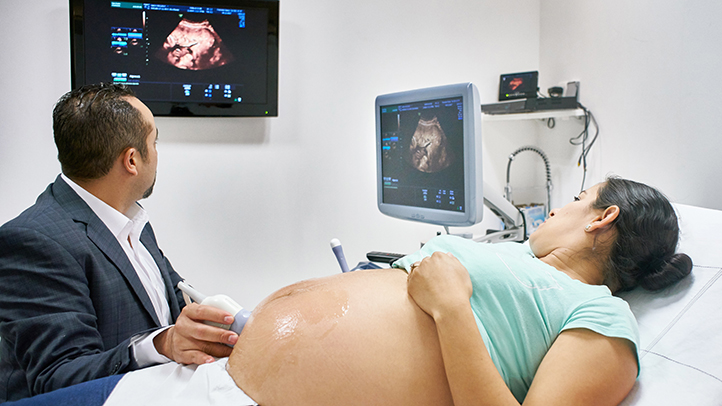
At Prabhu Women’s Hospital, we provide advanced 3D/4D sonography services that offer detailed imaging for pregnancy care and women’s health. With 3D sonography, patients get clear, high-resolution images, while 4D sonography adds real-time video, allowing parents to see their baby’s movements before birth. These technologies enhance diagnostic accuracy, support safe pregnancies, and create an emotional bonding experience for expecting families.
Clear and accurate imaging for pregnancy monitoring
Real-time 4D view of the baby’s movements and expressions
Improved diagnosis and enhanced parental bonding

How Does It Help in Women’s Health?
Sonography plays a vital role in women’s healthcare by monitoring pregnancy, detecting ovarian cysts, fibroids, and endometriosis, and guiding effective fertility treatments.
3D Sonography
4D Sonography
Services Tailored for You
Purpose of 3D-4D Sonography
Purpose of 3D-4D Sonography
Laparoscopy Services Tailored for You
Medical Diagnosis
Pregnancy Monitoring
Guidance for Procedures
Detecting Organ Damage
Evaluating Blood Flow
Assessing Soft Tissue Injuries
Non-invasive and Safe
Monitoring Treatment Progress
Evaluating Heart Function
What Are
Benefits of Sonography
Clear, high-definition images
Provides detailed 3D photos of your baby’s features, allowing for a more full glimpse than typical ultrasounds.
Real-time movement in 4D
Real-time video imaging allows you to watch your baby move, yawn, stretch, or grin, resulting in a wonderful bonding experience.
Safe and Non-invasive
Uses the same safe ultrasound technology as routine scans, which is perfectly hazardous to both mother and baby.
Improved diagnostic accuracy
Provides sharper pictures to help clinicians monitor foetal growth, position, and overall well-being with greater accuracy.
Stronger Emotional Connection
Seeing your baby’s face and movements strengthens emotional bonds among parents and family members.
Early Detection of Specific Conditions
Identifies physical anomalies or developmental concerns early on, allowing for more prompt medical planning.
Expert 3D/4D Sonography Imaging for Expectant Mothers
Our 3D/4D sonography services provide expectant parents with a clear, detailed view of their developing baby—capturing precious moments like smiles, yawns, and gentle movements in real time. These advanced ultrasound scans are completely safe, non-invasive, and emotionally rewarding. Using the latest imaging tools, our skilled sonographers deliver accurate assessments while creating a meaningful bonding experience for families. Whether you’re looking to connect with your baby or need detailed diagnostics, we ensure a warm, professional environment with expert care at every step
Process of 3D Laparoscopy
The ultrasound probe sends sound waves into the body, which bounce back to create 2D images of the baby and internal structures.
Advanced software combines multiple 2D images to form a detailed 3D image, showing clearer anatomy and facial features.
In 4D, these 3D images are continuously updated in real-time, allowing parents and doctors to see the baby’s movements live.
This technology not only helps doctors detect abnormalities with precision but also provides parents with an emotional bonding experience by allowing them to watch their baby in motion.
Book Your Sonography Consultation with Trusted Experts
Why Choose Prabhu Women's Hospital for 3D 4d Sonography?

Advanced 3D/4D Images
With 3D/4D sonography, doctors can find cleft lip, neural and bone problems early in the growth of an unborn baby.

Expert Medical Team
Expert doctors are using advanced sonography that can find fetal problems early by observing clear and updated images in real time.

Quality care, budget-friendly.
Modern sonography technology helps identify complications early by offering detailed and updated visuals.
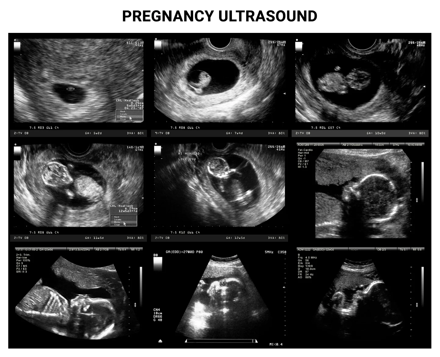
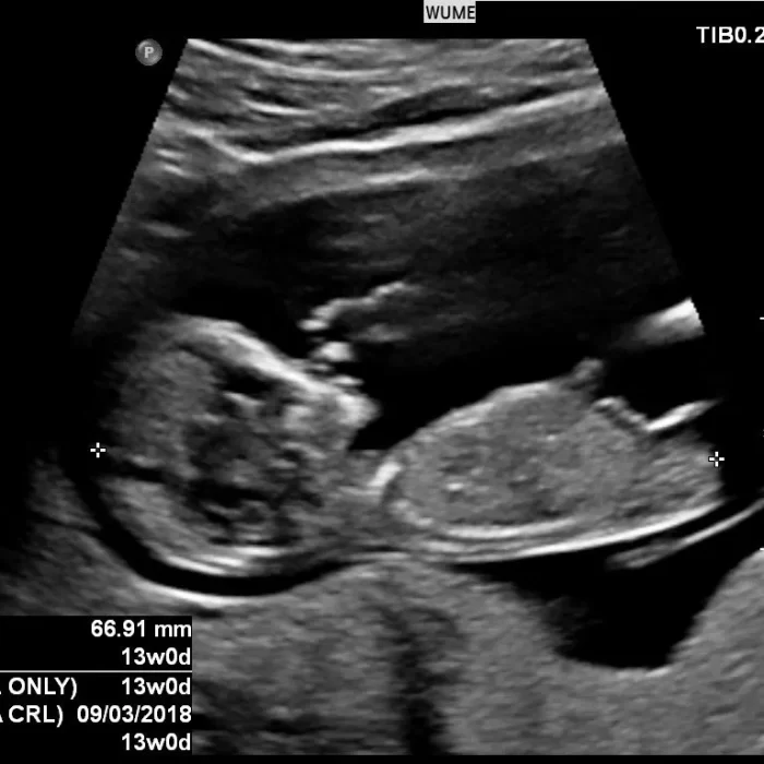
SONOGRAPHY GALLERY
See special moments of your baby captured through our sonography in this gallery


Frequently Asked Questions
What is the difference between 3D and 4D sonography?
3D sonography captures still, three-dimensional images of the baby, while 4D sonography adds the element of real-time motion—allowing you to see your baby move, yawn, stretch, or smile live on screen.
Is 3D/4D sonography safe for the baby and mother?
Yes. 3D/4D ultrasounds use the same safe ultrasound technology as traditional scans. They are non-invasive and have no known risks when performed by trained professionals.
When is the best time to get a 3D/4D ultrasound?
The best time is between 26 and 32 weeks of pregnancy. At this stage, there’s enough fat on the baby’s face for clearer images, and there’s still enough amniotic fluid to allow for good visualization.
Do I need a doctor’s referral for 3D/4D sonography?
While not always required, it’s best to consult your obstetrician before scheduling. In some cases, your doctor may recommend it for more detailed imaging or bonding purposes.
Can I bring family members for the scan?
Absolutely! We encourage you to share this special moment. Our comfortable scanning rooms are designed to let families experience the joy of seeing the baby together.
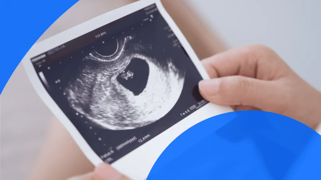
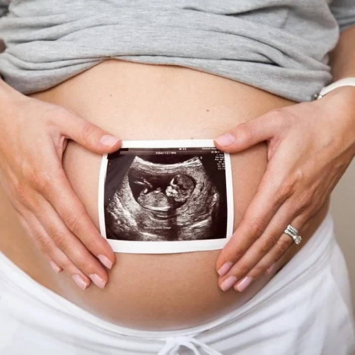
Will I receive photos or video of my baby?
Yes, we provide printed images and digital video clips (depending on the package) so you can cherish and share the experience with loved ones.